Mapping the distribution and quantity of melanin pigments in the skin can help doctors diagnose melanoma early, but pheomelanin — the pigment responsible for both the orange-reddishness in hair and a higher risk of hard-to-detect melanoma — is fairly difficult to detect in the skin.
However, researchers at Massachusetts General Hospital’s Wellman Center for Photomedicine have announced a breakthrough. It turns out that pheomelanin molecules vibrate unlike any other molecules in the body, allowing researchers to image the pigment using a special kind of microscopy technique.
Sam Osseiran, a scientist on the team led by Harvard University professor Conor Evans, will present the team’s findings April 2-5 at the OSA Biophotonics Congress in San Diego, California.
While eumelanin, the brown-black pigment found in most melanomas, is easily seen in the skin, the light-colored pheomelanin is much more difficult to detect. As a result, patients who have melanomas with this type of melanin are diagnosed at more advanced stages. Melanoma is the deadliest form of skin cancer, with more than 232,000 new cases and 55,000 deaths per year worldwide.
“There’s really no good way to see this mostly invisible pigment when it occurs in skin. So my team put our heads together, scouring for ways to see it,” Evans said in a press release. “We started to look through the Raman literature. Raman spectroscopy is a very mature technique that allows you to detect molecules by their unique chemical vibrations, which are themselves derived from the structure of the molecules.”
The team found that pheomelanin had a unique chemical structure — unlike any molecule in the body — so researchers assessed whether it also had a corresponding unique molecular vibration that could help image the melanin pigment.
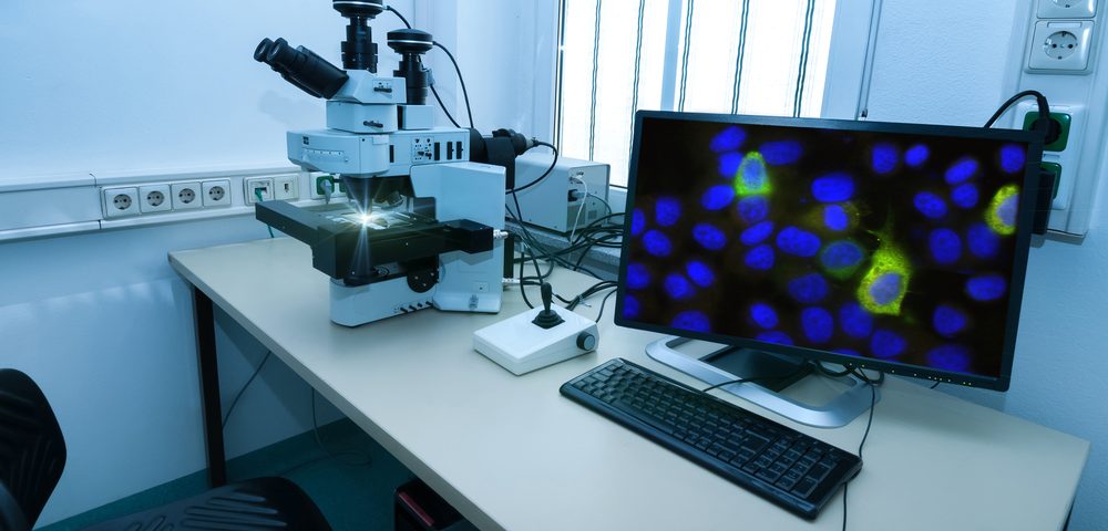
Using CARS microscopy, the team found that indeed it was the case. In CARS microscopy, two lasers focus on a sample, and the unique molecular vibration generates an image of the molecules. Osseiran said nobody had ever tried to do this before.
“We adjusted our system and aligned and tuned everything so that we could specifically target this one melanin pigment, pheomelanin,” he said. Coincidently, the team also found that a different form of microscopy — sum-frequency absorption (SFA) —could be used to detect both eumelanin and pheomelanin, allowing researchers to identify patients with high levels of these pigments in their skin.
“Sum-frequency absorption imaging allows you to visualize where all the melanin absorbers are within tissue,” said Evans. “As both CARS and SFA can be carried out at the same time, these two techniques can be used together to simultaneously image both melanin pigments.”
David Fischer, who first approached Evans to conduct the study, believes in the potential of these microscopy techniques to help diagnose melanoma patients early.
“This may offer a brand new tool for early diagnosis for some of the most lethal melanomas, possibly at a stage when they might still be curable,” said Fischer. “Time and time again, it is proven that early diagnosis saves lives.”


