 A new device allowing an early non-invasive examination of melanoma and other skin cancer lesions has been developed by a team of researchers from the University of Texas at Austin’s Cockrell School of Engineering.
The probe combines three different ways of using light to measure skin properties into one single device, thus allowing cancer detection without having to preform unnecessary biopsies.
The new skin cancer probe was described in a paper entitled “Design and characterization of a novel multimodal fiber-optic probe and spectroscopy system for skin cancer applications” and published in the Review of Scientific Instruments journal.
The common approach to diagnose skin cancer is to perform a biopsy, removing a piece of skin that shows a suspect lesion while preforming a histological analysis to detect cancer cells. However, biopsies are not 100% accurate, and for every detected case of skin cancer, about 25 negative biopsies are done, resulting in enormous monetary costs for the U.S. health care system and a distressing experience for the patient.
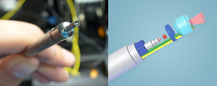
This cutting-edge probe integrates three common spectroscopic techniques — Raman spectroscopy, diffuse reflectance spectroscopy, and laser-induced fluorescence spectroscopy. This give the device the capacity to diagnose which skin lesions are more likely to be cancerous in a clear and precise way, therefore reducing the number and costs of negative biopsies and making it an improved screening tool for cancer. Each reading takes less than 5 seconds to perform and the probe itself is very small — about the size of a pen — and the computer equipment supporting it can fit inside a portable utility cart.
“As normal skin becomes cancerous, cell nuclei enlarge, the top layers of skin can thicken and the skin cells can increase their consumption of oxygen and become disorganized, altering the way light interacts with the tissue” study author James Tunnell, Associate Professor, Biomedical Engineering Department, University of Texas, told the American Institute of Physics.
To accurately detect these different changes would normally require multiple and separate spectroscopic techniques to detect absorption by proteins or vibrational modes of chemical bonds, such as those found in connective tissues, lipids, and cell nuclei.
The UT researchers are the first ones to combine all these different methods into a single probe that could be affordable enough to be widely used in the clinic.
“Skin is a natural organ to apply imaging and spectroscopy devices to because of its easy access. This probe that is able to combine all three spectral modalities is the next critical step to translating spectroscopic technology to the clinic,” added Dr. Tunnell.
The newly developed probe is already being tested in pilot clinical trials and the team has begun partnering with funding agencies and companies to eventually make the product available to the medical community.
